UA-38958768-1
Your browser version is outdated. We recommend that you update your browser to the latest version.

The Importance of the Thoracic Spine in Shoulder Mechanics

Posted 9/28/2013

By Chris Feil, DC and William E. Morgan, DC

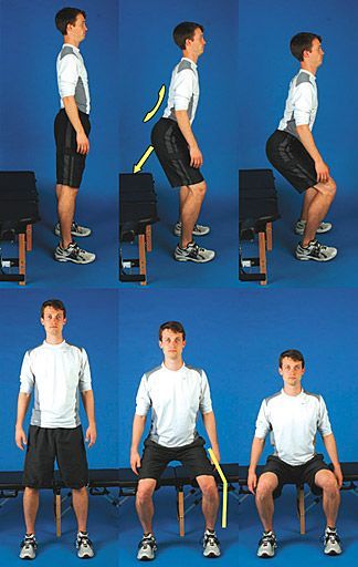
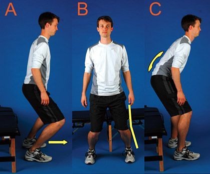
As discussed in previous articles in this series, cross-fitness programs tend to advocate exercises that require maximal shoulder end-range motions: pull-ups (often with a kip or a jerk motion at end range), handstand presses, press squats, push-presses, clean and presses, kettlebell overhead lifting, gymnastic ring work and other similar exercises.

To perform these motions without injury requires unfettered shoulder range of motion and optimum shoulder stability. While an argument may be made that these exercises help to create coordinated athletic patterns of strength, agility, and stamina, they still have a strong potential for causing shoulder injuries. Often not included in the explanation of the workout of the day in cross-fitness programs is the need for the participant to have flawless shoulder mechanics and strength.

Impingement of the rotator cuff muscle (supraspinatus) develops when the space between the rigid coracoacromial arch and the head of the humerus narrows. The muscles and tendons of the cuff that pass through this space begin to fray and eventually may tear because they are pinched between these hard surfaces. Why does this space narrow? Anatomically, it narrows due to bony spurs, degenerative changes, or soft-tissue thickening. Functionally, the space narrows due to dysfunctional synchronicity of the rotator cuff muscles, aberrant scapular-humeral rhythm, or faulty scapular positioning during overhead arm movements.

To maintain the subacromial space in an overhead arm movement, the scapula must retract and tilt posterior.7 A shortened pectoralis minor will cause the scapula to tilt anterior, contributing to a functional shoulder impingement. A study conducted at Ohio State University found that subjects with tight and short pectoralis minor muscles displayed similar scapular kinematics as individuals with shoulder impingements.1 Scapular retraction is affected by the mobility of the thoracic spine and rib cage upon which it glides.9

Thoracic Spine Mechanics and Shoulder Pain

Shoulder Reach 
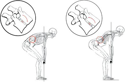
Normal scapular and thoracic spine motion allows optimal mechanics for athletic shoulder motions (left). Increased thoracic kyphosis, reduced thoracic mobility, or scapular protraction from pectoralis minor tautness can contribute to shoulder impairment and injury (right). The concept of scapulo-humeral rhythm is well-documented,4,7 and the rhythm is fundamental to maintaining the subacromial space. The long-accepted linear ratio of scapular rotation to arm motion in adduction is 1:2,2 though the true interaction of the scapula and arm is not linear, but more curvilinear in nature.8 There is very little scapular rotation in the first 60 degrees of arm abduction, and then the scapula progressively begins to rotate as the arm travels to a full overhead position.4

Normal thoracic-humeral rhythm is important for injury prevention. Thoracic mobility becomes increasingly important in athletic overhead activities. The higher an athlete raises their arm, the more thoracic motion is needed from the thoracic spine to maintain the proper relative shoulder alignment. Individuals with a shoulder impingement have statistically less thoracic mobility and a more kyphotic thoracic spinal posture than individuals with healthy shoulders.3,6-7 A few weeks of cross-fitness training with a rigid thoracic spine could lead to injury and impairment of the rotator cuff.

Does T4 Syndrome Exist?

T4 syndrome has been accepted as fact by many clinicians over the past decade, in spite of a lack of evidence to support its existence. The theory of T4 syndrome attributes many of the problems seen in shoulders to a loss in the normal extension that takes place at the T4 vertebra.5 We should note that T4 syndrome has also been credited with causing diffuse arm, shoulder, and torso pain and sensory symptoms.

A review of the literature review did not find research substantiating association of the T4 syndrome to shoulder function. However, we did find a boatload of seminar notes elevating the "T4 syndrome" to a height not supported by the current body of research. T4 syndrome is based on clinical antidotal experience rather than scientific evidence. While there is no substantial evidence that T4 syndrome directly relates to shoulder impingement, there is some current research pointing to the lower thoracic spine as being a fundamental component of shoulder motion.4

If accurate, this research suggests that the majority of thoracic extension occurs in the lower thoracic spine during overhead arm movement in asymptomatic shoulders, especially with both arms in an elevated position, as is seen in an overhead squat or overhead press.

In the same study, upper thoracic extension was present in full overhead arm motion, but it was not deemed to be a significant variable in shoulder function.4 However, what the study did find statistically significant in the upper thoracic spine was lateral bending and ipsilateral rotation during single-arm elevation. Theodoridis and Ruston also found this repeatable relationship of overhead arm movement causing an ipsilateral coupling pattern between lateral flexion and rotation, which was repeatable and comparable for both arm elevation planes in healthy subjects.11

Thoracic Joint Manipulation

It appears that simply assessing T4 extension in a cross-fitness athlete may miss significant dysfunctions in the thoracic spine that affect the thoracic-humeral rhythm. For example, optimal shoulder function in a single-arm kettelbell swing may be improved by use of manual therapy when specifically addressing lateral bend and rotation components of the upper thoracic spine and any loss of extension in the lower mid-thoracic spine.

Thoracic joint manipulation might be the simplest answer to reducing pain in an impinged shoulder. In a 2009 study by Strunce and colleagues, a thoracic spinal manipulative thrust was performed on a sample of 56 individuals with symptomatic shoulders from impingement. After two days, there was a significant decrease in pain levels in over 50 percent of individuals.10

Before giving a list of boring rotator-cuff strengthening exercises to a cross-fitness athlete who is performing fun, dynamic exercises in their daily workouts, remember that a key component to overhead arm motion is thoracic mobility. When the alignment of the shoulder and the subacromial space is compromised from the loss of thoracic spine mobility and/or muscle tightness, it doesn't matter what tension of rehab exercise band is used until the underlying cause of impingement is addressed. Evaluation and appropriate treatment of the spine should be considered when shoulder dysfunction is present.

The cross-fitness emphasis on pull-ups and overhead lifting may produce a glut of shoulder injuries from otherwise dormant thoracic and shoulder impairments. By recognizing the functional relationship between the thoracic spine and the shoulder joints, we can help athletes remain active and pain-free as they engage in their preferred activities.

References

  1. Borstad JD, Ludewig PM. The effect of long versus short pectoralis minor resting length on scapular kinematics in healthy individuals. Journal of Orthopedic Sports Physical Therapy, 2005;4:227-238.
  2. Codman EA. The Shoulder; Rupture of the Supraspinatus Tendon and Other Lesions in or About the Subacromial Bursa. Thomas Todd, Boston, 1934.
  3. Crawford HJ, Jull GA. The influence of thoracic posture and movement on range of arm elevation. Physiotherapy Theory and Practice, 1993;9:143-148.
  4. Crosbie J, Kilbreath SL, Hollmann L, York S. Scapulohumeral rhythm and associated spinal motion. Clinical Biomechanics, 2008;23:184-192.
  5. DeFranca GG, Levine LJ. The T4 syndrome. Journal of Manipulative and Physiological Therapeutics, 1995;1:34-37.
  6. Greenfield B, et al. Posture in patients with shoulder overuse injuries and healthy individuals. Journal of Orthopedic Sports Physical Therapy, 1995;5:287-295.
  7. Kibler BW. The role of the scapula in athletic shoulder function. The American Journal of Sports Medicine, 1998;26:325-337.
  8. McClure PW, Michener LA, Sennett BJ, Karduna AR. Direct 3-dimensional measurement of scapular kinematics during dynamic movements in vivo. Journal of Shoulder and Elbow Surgery, 2001;10:269-277.
  9. Stewart S, Jull GA, Ng, JKF, Willems JM. An initial analysis of thoracic spine movement during unilateral arm elevation. Journal of Manual & Manipulative Therapy, 1995;3:15-20.
  10. Strunce J, et al. The immediate effects of thoracic spine and rib manipulation on subjects with primary complaints of shoulder pain. Journal of Manual & Manipulative Therapy, 2009;17:230-236.
  11. Theodoridis D, Ruston S. The effect of shoulder movements on thoracic spine 3D motion. Clinical Biomechanics, 2002;17:418-421.

The views expressed in this article are those of the authors and do not necessarily reflect the official policy or position of the Department of the Navy, Department of Defense or the U.S. government. This is the fourth in a continuing series on cross-fitness training. The first article appeared in the Sept. 9, 2009 issue; the second in the Oct. 7, 2009 issue; the third in the Nov. 4, 2009 issue; and the fourth in the Jan. 29, 2010 issue.


Reprinted from Dynamic Chiropractic – May 6, 2010, Vol. 28, Issue 10


Read the rest of this entry »

Functional Integrity of the Pelvis & Hips

Posted 9/28/2013

Gluteal Activation Enhances Athleticism and Injury Prevention

By Chris Feil, DC and William E. Morgan, DC

Editor's note: This is the fourth in a continuing series on cross-fitness training. The first article appeared in the Sept. 9, 2009 issue; the second in the Oct. 7, 2009 issue; and the third in the Nov. 4, 2009 issue.

For most athletes, success is largely dependent on optimal functioning of the gluteal muscles (gluteus maximus, medius and minimus), and functional integrity of the hips and pelvis. Unfortunately, functional training and evaluation is not well-understood by many practitioners and athletes. Appropriate gluteal activation and pelvic-hip control is not only important to the rising number of cross-fitness devotees for generating maximal athletic power; it is also important to virtually every chiropractic patient. In addition to generating athletic power, proper hip function is valuable in the prevention of injuries to the knees, hips, pelvis and lower back. The cross-fitness activities of squatting, cleans, kettlebell swings, tire flipping, medicine ball tossing, and sprinting are all multi-joint movements that require hip involvement. Let's discuss methods to maximize proper hip motion and form during these activities.

Many in modern society have what Stuart McGill, PhD, calls "gluteal amnesia."1 Dr. McGill has identified that when athletes [or any of us] lose the ability to engage our hips during athletic activities or exercises (such as cross-fitness programs), this adversely affects performance and increases the likelihood of injury. What Dr. McGill calls "gluteal amnesia," we might identify as loss of functional hip integrity: essentially the loss of the normal volitional ability to move one's hips through their range of motion with appropriate muscle activation.

In addition to muscular inhibitions, other factors that may contribute to motion dysfunctions are soft-tissue contractures or restrictions and articular fixations. While chiropractic adjustments may directly affect these restrictive lesions, knowledge of gluteal activation is also required to teach patients how to properly activate these muscles.

Function of the Gluteus Maximus

The gluteus maximus (GM) is the largest muscle of the body, and it is the major driver in lifting, throwing, swinging, pushing and running (particularly when sprinting and running hills). The GM originates at the crest of the pelvis, the dorsal sacral ligament, along with some fibers that originate from the thoracodorsal fascia. The [distal] insertions attach to the femur and to the iliotibial band. The GM helps stabilize the sacroiliac (SI) joints by causing force closure, essentially forming a self-bracing, protective compressive force to maintain the alignment and reduce shear forces on the SI joint. The GM attachment to the sacral ligaments may aid in pelvic stability due to active ligament tightening by gluteal contraction.2

The late professor Vladimir Janda associated decreased GM control in his theory of lower cross syndrome.3-4 This muscle imbalance is associated with the pelvic posture variation called anterior pelvic tilt. Observing anterior pelvic tilt during a postural examination of a patient should provoke you to further screen for gluteal dysfunction.

The gluteal muscles are the primary extensors of the hip, but hip extension is only part of their role in true athletic function. The muscle fibers of the GM are oriented diagonally, sloping laterally and caudally from their origin. With this orientation, the GM muscles contribute to external rotation and abduction of the thigh. Both hip extension and thigh external rotation are the "concentric" motions of the GM muscle.

It is important to remember that muscles have two contractile functions: concentric contractions (muscle shortening) and eccentric contractions (muscle lengthening). Eccentric muscle contraction is actually more powerful and more efficient than concentric contractions. The eccentric function of the GM muscle will limit and control hip flexion and thigh internal rotation.

Because of its fiber orientation, the GM serves as a primary muscular shock absorber for the hip and knee joint. Just as the hydraulic shock compresses and dampens the load in a car, the GM dissipates the forces in the athletic movements of jumping, landing, and lateral agility motions by eccentrically absorbing forces and limiting movements endangering joints of the lower extremities.5 The link between GM dysfunction and uncontrolled valgus and internal rotation motions of the knee has particular clinical significance and will be discussed in greater detail in an upcoming article.

Tremendous loads can be transmitted through the acetabulofemoral joint if the force is not dampened by the adjacent muscles. This shock absorption function is important to understand in a culture in which so many degenerative hip disorders occur. As many desk-bound workers with inadequate GM control participate in cross-fitness programs, they are unknowingly placing themselves at greater risk for osteoarthritis in their hips.

Squat Functional Screen: Passing Figure 1: Squat Functional Screen: Passing. Normal lumbar lordosis, posterior travel of hips, varus knee position, and limited anterior travel of knees. Initiating Movement at the Hip and GM Activation

Elite power-lifters are able to squat more than 1,000 lbs injury-free through very purposeful activation of the GM and maximizing hip motion with the hip hinge.6 The term hip hinge refers to truncal motion in which the lumbar spine is fixed in a neutral lordosis and all motion occurs at the acetabular joint, not the spine.

An ideal squat begins by securing the toes and heels firmly on the floor. The lumbar spine should be fixed or "locked" into a neutral lordosis throughout the squatting motion. While descending, the buttocks should travel back and down; using a stool, gym ball, or chair as a target may be beneficial in learning this motion pattern. A wide stance is preferred, and the participant should be mindful not to allow the knees to travel forward.

Purposely engage the GM throughout the squatting motion both during decent and ascent; the use of an elastic exercise band around the thighs will help the patient to consciously engage the GM. The ascent phase of a squat reverses the motion groove of the descent phase. It should also be noted that the patient needs to stiffen the core during hip hinges and squatting

The Squat as a Functional Screen for Gluteal Activation

Inspecting a patient's ability to squat is a practical method for clinical screening of the lumbo-pelvic-femur chain. While the patient performs repetitive squatting motions, analyze the three main components of gluteal involvement: hip extension, flexion and external rotation.


Squat Functional Screen - Failures

Figure 2: Squat Functional Screen - Failures. Anterior knee travel, valgus knee position, and loss of lumbar lordosis.

A key point in assessment is the initiation of hip movement before knee flexion or ankle dorsiflexion. The knee should remain in a varus position throughout the squatting motion. The lumbar lordosis should remain unchanged during the entire squat. (Figure 1) Early anterior knee translation (Figure 2a), a valgus knee angle (Figure 2b), and a flexed lumbar spine (Figure 2c) are failures for the squat functional screen.

Squat Functional Screen - Palpation Figure 3: Squat Functional Screen - Palpation. The clinician provides challenge by applying medially directed pressure on the knees while the patient performs a squat. The examiner should palpate the GM for activation throughout the movement of both ascending and descending phases of the squat. In addition to palpating the GM, activation of the gluteus maximus can be tested by applying medial pressure to the knees. If the GM is engaged the examiner will note springy, firm resistance. With proper GM function, it should be difficult for the examiner to push the squatter's knee into a valgus position. (Figure 3) With practice and patience, you will be able to identify motion defects and provide precise recommendations for improving motion patterns.

Athletes performing cross-fitness feats of strength, agility, and endurance with functional impairments can expect to have reduced levels of performance and increased occurrences of injury and infirmity. Gluteal activation and properly functioning hip mechanics are fundamental components of proper motion and maximized athletic performance. Cross-fitness devotees with impaired gluteal/hip function can expect diminished performances and increased risk of injury. An astute clinician should be able to observe a cross-fitness athlete's squat and discern gluteal function and activity, correcting those at risk before injury occurs.

References

  1. McGill S. Low Lack Disorders: Evidence-Based Prevention and Rehabilitation, 2nd Edition. Champaign: Human Kinetics, 2007:110-112.
  2. Wilson J, et al. A structured review of the role of gluteus maximus in rehabilitation. New Zealand Journal of Physiotherapy, 2005;33(3):95-100.
  3. Bullock-Saxton JE, Janda V, Bullock MI. Reflex activation of gluteal muscles in walking. Spine, 1993;18(6):704-8.
  4. Morris CE, Chaitow L, Janda V. Functional Examination for Low Back Syndromes. In: Morris C. Low Back Syndromes: Integrated Clinical Management. McGraw-Hill, 2006:347.
  5. Boling MC, Padua DA, Creighton RA. Concentric and eccentric torque of the hip musculature in individuals with and without patellofemoral pain. Journal of Athletic Training, 2009;44(1):7-13.
  6. Liebenson C. The hip hinge. Journal of Bodywork and Movement Therapies, 2003;7:151-152.

The views expressed in this article are those of the authors and do not necessarily reflect the official policy or position of the Department of the Navy, Department of Defense or the U.S. government.


This is a reprint from Dynamic Chiropractic – January 29, 2010, Vol. 28, Issue 03


Read the rest of this entry »

Core Stiffness There is No Fitness

Posted 9/28/2013

Without the Stiffness There Is No Fitness

By William E. Morgan, DC and Chris Feil, DC

Editor's note: This is the third in a continuing series on cross-fitness training. The first article appeared in the Sept. 9 issue; the second in the Oct. 7 issue.


The rising tide of cross-fitness popularity, for all of its potential fitness benefits, has the potential to cause a tsunami of lumbar spine injures if improperly implemented. In this third installment in our series on cross-fitness, let's discuss the importance of core muscle activation during cross-fitness exercise. Much has been written about the role of the core muscles in protecting the spine from injury in recent years, but this information is taking too long to trickle down to the gyms and cross-fitness clubs around the world. Properly engaging the core can enhance athleticism and reduce the risk of injury when performing rigorous exercise.   

The Core

Muscle Direction Figure 1: The posterior/superficial fibers of the thoracolumbar fascia angle up and away from the spine. Though these fibers are continuous with the latisimus dorsi, they are connected to the transverse abdominis (TrA) through the lateral raphe (based on Bogduk11). The term core is bantered about in all corners of the health and fitness industry, yet to most people who use this word it remains a vague, almost nebulous description of supportive stomach and back muscles. We would like to solidify the term and provide a workable definition of the core, at least as we currently understand the term: The core muscles are the truncal muscles that support and stabilize the torso, protect the spine, and allow power transfer through the torso.

The core includes global stabilization muscles such as the transverse abdominis (TrA), the internal oblique (IO), and external oblique (EO); and intersegmental stabilizers such as the multifidus muscles.1 Since the list of muscles composing the core is a moving target depending on authorship, we would rather concentrate on core function than try to generate a composite list of the core muscles.  However, certain muscles are central to any discussion of the core: the TrA, OI, OE, multifidus, rectus abdominis (RA), quadratus lumborum (QL), and the muscles of the erector spinae. Some would add the iliopsoas, latisimus dorsi, gluteal muscles, hip adductors, and hamstrings to this list.

The individual muscles of the core are each capable of contributing to several different functions, and no function is isolated to an individual muscle. The core is a complex and integrated network of muscles that work in synchrony to support the torso with stiffness and strength. It is impractical to try to isolate the function of individual muscles such as the TrA. In fact, when the core muscles stiffen in concert, their total strength surpasses the sum of the individual muscles.

Muscle Cross Section Figure 2: The anterior/deeper fibers of the thoracolumbar fascia angle downward and away from the spine from L2- L5. They are also connected to the TrA and the internal oblique (IO) by means of the lateral raphe (based on Bogduk11). In recent years, there has been a movement afoot that promotes abdominal hollowing,1 pulling in the abdominal muscles in an attempt to isolate the TrA and indirectly activate the multifidus muscles. Stuart McGill, PhD, has found that core bracing is superior to abdominal hollowing in regards to protecting the spine from injury. Vera-Garcia (with McGill as a co-author) found that hollowing actually inhibits the multifidus' response to perturbation,2 actually reducing core stabilization.

Your Internal Weight-Lifting Belt

Weight-training belts are no longer the rage they once were and their use is ebbing in most of the realm of physical culture. This is due in part to the knowledge that weight-lifting belts are not necessary. One study revealed that the advantage in using belts may come from perceived rather than actual protection and performance enhancement.3 Belts essentially increase the amount that lifters are willing to lift. Belts interfere with the natural intrinsic stabilization of the trunk without substantial benefit and should generally be avoided.4-9

Thoracolumbar Figure 3: When the TrA and IO contract, the anterior and posterior layers of the thoracolumbar fascia are pulled taut, approximating the spinous processes [from L2-L5], and a stiffening effect takes place (based on Bogduk11). We each possess a natural, built-in weight belt that is activated by the muscles of the core. Your intrinsic stabilization corset consists of a combination of the TrA and the thoracolumbar fascia. The posterior layer of the thoracolumbar fascia angles up away from the spine (Figure 1), whereas the anterior layer of the thoracolumbar fascia angles down and away from the spine (Figure 2). They both are joined to the TrA by the lateral raphe. So, when the TrA stiffens, the contraction produces a Poisson's effect,10 which causes the spinous processes to approximate in a protective manner (Figures 3 and 4).11

A collateral benefit of TrA contraction is activation of the multifidus muscle.12 The multifidus provides intersegmental stabilization through its stiffening effect, but is difficult to contract voluntarily. Together, the global and intersegmental stabilizers protect the spine from excessive shear and torsional forces.13

Bracing the Core With Muscular Stiffness

Learning to brace the core is an important component in protecting the spine and enhancing athletic performance. While many athletes intuitively brace their core during athletic exertion, others require training. It takes only a few minutes to learn how to coax the core muscles into a protective stiffened brace, but may take months to imbed a permanent neurological groove of bracing into a particular athletic motion pattern.

Thoracolumbar Fascia Figure 4: This schematic amplifies the concept of Poisson's effect. The contraction of the transverse and oblique abdominal muscles pulls on the lateral raphe, which produces a mild approximating tension of the lumbar spinous processes. Begin in a relaxed standing posture; place the fingertips of one hand on the lumbar paraspinal muscles just to the side of the spinous processes. The other hand should be positioned on the abdominal muscles at the level of the ASIS. Bend at the waist until you feel the muscles of your lower back contract under the fingers on your back. Note how this feels and then arch your spine until the spinal muscles relax under your fingers. While maintaining this position, stiffen the abdominal muscles as if you were about to be punched in the gut. You should feel your spinal muscles contract like they did when you bent over at the waist. Note how this feels with both hands. Practice engaging these muscles until it takes little conscious effort.

If bracing in this manner aggravates a spinal condition, reduce the degree of abdominal contraction. Maximal stiffness is not required. Practice in the range of 10 percent to 25 percent of maximal stiffening. Stiffening should accompany strenuous athletic exertion. When establishing neurological groove patterns for compound motor patterns, make sure to include bracing. For squatting motions, stiffen the core throughout the entire motion, even when no weight is used.

A dilemma that often accompanies core stiffening exercises is interference with diaphragmatic breathing. When first learning to stiffen the core, consciously engage in diaphragmatic breathing until it becomes routine. Once core bracing and diaphragmatic breathing are mastered, they should be practiced or rehearsed while in exertion-induced respiratory distress until it becomes natural. Practice performing cardiovascular interval training while concentrating on core bracing and diaphragmatic breathing. In time, these two activities will be imbedded in your neurology to the point of not requiring conscious intercession.

Core Stiffness Is Fundamental to Cross Fitness

Core bracing fits into one of the creeds of the cross-fitness movement: integration of compound motion patterns rather than muscle isolation. A properly functioning and reactive core is required for high levels of athleticism whether you are an elite athlete, a cross-fitness devotee, or even an average weekend golfer. Certainly cross-fitness injuries will be curtailed if athletes maintain proper form and utilize core bracing when performing athletic activities such as squatting, tire flipping, dead-lifting, plyometric jumping drills, kettlebell drills and agility drills. If our patients are going to engage in cross-fitness programs, it is our duty to prepare them properly through treatment, prevention and education.

References

  1. Richardson C, Jull G,  et al. Therapeutic Exercise for Spinal Segmental Stabilization in Low Back Pain. Churchill Livingstone, Edinburgh, 1999:22-25.
  2. Vera-Garcia FJ, Elvira JL, Brown SH, McGill SM. Effects of abdominal stabilization maneuvers on the control of spine motion and stability against sudden trunk perturbations. J Electromyogr Kinesiol, 2006 Sep 20;17(5):556-67.
  3. McCoy MA, Congleton JJ, Johnston WL, Jiang BC. The role of lifting belts in manual lifting. Int J Ind Ergonomics, 1988;2:259-266.
  4. Majkowski GR, Jovag BW, Taylor BT, Taylor MS, Allison SC Stetts DM, Clayton RL. The effect of back belt use on isometric lifting force and fatigue of the lumbar paraspinal muscles.Spine, 1998;23(19):2104-2109.
  5. National Institute for Occupational Safety and Health. Workplace Use of Back Belts: Review and Recommendations. Rockville, MD: Department of Health and Human Services (National Institute of Occupational Safety and Health), Publication No. 94-122, 1994.
  6. Mitchell LV, Lawler FH, Bowen D, Mote W Asundi P, Purswell J. Effectiveness and cost-effectiveness of employer-issued back belts in areas of high risk for back injury. J Occup Med, 1994 Jan;36(1):90-94.
  7. Thomas JS, Lavender SA, Corcos DM, Andersson GB. Effect of lifting belts on trunk muscle activation during a suddenly applied load. Hum Factors, 1999 Dec;41(4):670-6.
  8. Reyna JR, Leggett SH, Kenny K, Holmes B, Mooney V. The effect of lumbar belts on isolated lumbar muscle strength and dynamic capacity. Spine, 1995;20(1)68-73.
  9. McGill SM, Norman RW, Sharratt MT. The effect of an abdominal belt on trunk muscle activity and intra-abdominal pressure during squat lifts. Ergonomics, 1990 Feb;33(2):147-60.
  10. See www.ecourses.ou.edu/cgi-bin/view_anime.cgi?file=m1421.swf&course=me&chap_sec=01.4
  11. Bogduk N. Clinical Anatomy of the Lumbar Spine and Sacrum, 3rd Edition. Churchill Livingstone, Edinburgh, 2001.
  12. Hides JA, Jull GA, Richardson CA. Long-term effects of specific stabilizing exercises for first-episode low back pain. Spine, 2001;26:E243-8.
  13. Moseley GL, Hodges PW, Gandevia SC. Deep & superficial fibers of lumbar multifidus are differentially active during voluntary arm movements. Spine, 2002;27(2):E29-36.

The views expressed in this article are those of the authors and do not necessarily reflect the official policy or position of the Department of the Navy, Department of Defense or the U.S. government. This is the third in a series of articles addressing cross-fitness. Future articles will present foundational components of core stabilization and gluteal activation, as well as the prevention of shoulder injuries.

Reprinted from Dynamic Chiropractic – November 4, 2009, Vol. 27, Issue 23

Read the rest of this entry »

Cross-Fitness Injury Prevention: Protecting the Lumbar Disc In Squatting Motions

Posted 9/28/2013

By William E. Morgan, DC and Chris Feil, DC

Editor's note: This is the second in a continuing series on cross-fitness training. The first article appeared in the Sept. 9 issue.

With the rising number of people joining the cross-fitness movement and the lack of adequately trained coaches, there is a high likelihood that we will continue to experience an increase in cross-fitness-related injuries.

While the cross-fitness movement has many positive benefits and we do not want to discourage people from exercising, we need to be able to protect those interested in this increasingly popular exercise program.

Cross-fitness, as stated in our previous article, is based largely on the implementation of compound motion patterns that use groups of muscles, rather than isolated muscles. The workouts are varied and seek to maintain physiological confusion and turbulence to continually challenge participants' bodies to adapt to higher levels of fitness. A mainstay of these programs is squatting motions.

Our first article provided an overview of this fitness phenomenon. Now let's discuss some specific biomechanical considerations of the exercises commonly performed in cross-fitness training, with specific emphasis on the principles of protecting the lumbar disc while squatting.

Perfect Practice Makes Perfect; Practice Just Makes Permanent

Squatting is required to perform dozens of exercise variations used in cross-fitness programs: squats, dead-lifting, power cleans, clean and jerks, truck tire flipping, kettlebell presses, bends and thrusts (aka, burpies), lunge walking, box jumping, jumping lunge squats and more. These exercises are frequently performed rapidly while striving to execute a high number of repetitions as part of a timed workout. Exercising in this manner quickly fatigues the participants, increasing the likelihood of form degradation. This is usually manifested in flexion of the lumbar spine.

We know from Alf Nachemson's work1 that curling the lumbar spine forward increases disc pressure, especially when lifting. We also know that lumbar flexion encourages migration of the nucleus pulposa posterior toward the spinal nerves (Figure 1). Maintenance of normal lumbar lordosis is protective for preventing disc injuries (Figure 2).


image

Figure 1 (left): This lifter curls their spine forward. This method of lifting causes the nucleus of the disc to migrate posterior, increasing the likelihood of a lumbar disc derangement. This method of lifting is dangerous and should be avoided. Figure 2 (right): Maintenance of a normal lumbar lordosis when squatting is protective for preventing disc injuries. Note that this lifter bends forward pivoting only at the hips, not the spine. In this manner, forward truncal fl exion can safely take place and lumbar disc injury is avoided.  

It is important to maintain the lumber spine in lordosis whenever squatting or bending. The lower you bend, the more likely you are to flex the spine. The more you flex your spine, the more likely you are to sustain a disc injury. Truck tire flipping and sandbag lifting, both of which are popular cross-fitness exercises, require squatting deep enough to allow the back of your hand to reach the ground prior to lifting. This is more dangerous than dead-lifting a barbell that is 10-12 inches above the floor. Rapid tire flipping while competing against a stopwatch further increases the likelihood of injury.

Proper squatting technique will reduce the likelihood of spine injury. The primary components of a proper squat include: maintaining lumbar lordosis, proper hip motion, gluteal activation, and core stiffening. These are all integral parts of the squatting motion and are difficult to tease out into individual components. For the sake of clarity, we will address each component in separate articles while alluding to the other components.

It is our contention that before anyone begins a cross-fitness program, they should know how to perform a proper squatting motion, and have practiced it enough times to establish an enduring neurological "groove" - a neurological motor pattern that has been practiced to the point of becoming the dominant sequence of motion for a particular action. In regards to neurologic motor patterns, practice does not make perfect. Perfect practice makes perfect. Practice just makes permanent.

Cross-fitness aspirants should practice proper squatting to the point that they virtually cannot squat wrong. Only after a safe squatting groove is established should a participant progress to more advanced programs.

The Proper Squat

Begin by locking the lumbar spine into natural lordosis while standing with your feet slightly wider than shoulder-width apart and your toes turned out slightly. The chest should be held out and the abdominal muscles stiffened. Continue to hold the lumbar spine in a stiff lordosis and use the movement of your hips to lower your body. Your buttocks should travel inferior and posterior as you squat.

Fig 3 Figure 3: An effective method to enforce proper squatting technique is the use of a rod held against the spine as a proprioceptive feedback tool. If the subject varies from proper technique, they will feel their back change positions in relation to the rod. It cannot be emphasized enough that hip motion is more important than spine motion when it comes to protecting the lumbar spine. Virtually all lumbopelvic motion of squatting should come from the hips. As you descend and ascend during the squat, attempt to engage your gluteal muscles by maintaining a constant low-level contraction of external rotation of the hips. (We will discuss gluteal activation more fully in a subsequent article.)

An effective method to enforce proper squatting technique is the use of a rod held against the spine as a proprioceptive feedback tool (Figure 3). If the subject varies from proper technique, they will feel their back change positions in relation to the rod. When done correctly, the spine will maintain the same contact with the rod throughout the entire squatting motion and the hips will do virtually all of the movement.

In addition to proper technique, it is important to identify the most hazardous time of day to challenge the lumbar discs. Due to the diurnal effects of disc hydration, discs hydrate at night and lose hydration throughout the weight-bearing hours of the day; discs are larger and more prone to injury in the early morning. For this reason, we recommend that strenuous activities such as rigorous squatting exercises be avoided for the first 1 1/2 hours after rising.


Componets of Proper Squatting

   Stiffening of the core muscles
   Maintain a lumbar lordosis
   Use your hips, not your spine
   Gluteal activation

Whether you are an 85-year-old woman with osteoporosis or the most avid cross-fitness devotee, proper squatting technique is important.  Learning how to squat safely should be part of virtually every patient's program of spinal hygiene. Squatting is not just an exercise; it is an integral part of human existence and is important for independent living. (One of the factors that will compel the elderly or infirmed to relocate into an assisted living community is the inability to squat onto a commode or to rise from it.) Aside from helping cross-fitness enthusiasts, your increased knowledge of the technicalities of squatting motions should help virtually all of your patients enjoy healthier and more fulfilling lives.

Reference

  1. Nachemson AL. In vivo discometry in lumbar discs with irregular nucleograms. Acta Orthop Scand, 1965;36;426.

The views expressed in this article are those of the authors and do not necessarily reflect the official policy or position of the Department of the Navy, Department of Defense or the U.S. government. This is the second in a series of articles addressing cross-fitness. Future articles will present foundational components of core stabilization and gluteal activation, as well as the prevention of shoulder injuries.


Reprinted from Dynamic Chiropractic – October 7, 2009, Vol. 27, Issue 21

Read the rest of this entry »

The Cross-Fitness Craze

Time is Tissue

Building a Culture of Continuous Improvement

Posted 9/26/2013

Self-Evaluation of Practice Performance


In institutional healthcare there is a requirement of continual self-reflection and improvement. Any chiropractor who has integrated into an institutional healthcare facility is probably already familiar with the term P.E. & I. –Performance (or Process) Evaluation and Improvement.   Accrediting bodies in mainstream healthcare require ongoing projects that include self-reflection, analysis and improvement for hospitals and medical centers. In fact some medical societies and organizations are also implementing P.E. & I. as a requisite to certification.


If you are entering into integration with an institutional health care faculty (hospital-based, university-based, military, VA, public health service clinic…) you should be familiar with this method of continual analysis and improvement.  In fact, even if you are not involved in an organization that requires ongoing P.E. & I. projects, it is a virtuous discipline to incorporate into your practice.


The concept is simple:  Identify an area in your clinic that needs improvement, select a metric (unit of measurement), measure existing trends using this metric, implement a program of improvement, re-measure metric, modify program as required to maximize your improvement.


Some medical specialties, such as the American Board of Internal Medicine, have created kits for practice evaluation and improvement.  A physician completing these projects can even receive CE credits.


An Example of Performance Evaluation and Improvement in Action


Selecting the Process to be Improved and the Measurement Metric

A doctor is concerned about her utilization of outcome measurement tools in her clinical practice.  For her metric she decides to measure the number of lower back pain patients completing Roland-Morris questionnaires.  


Collecting Initial Data

The doctor has her staff randomly survey 100 patient charts and finds out that 30 % of her patients with lower back pain have completed these surveys on their initial visits, and 5% complete follow-up questionnaires.  


Designing and Implementing a Corrective Plan


She selects a plan that empowers her staff to have all new lower back pain patients complete a Roland-Morris at the time of their first visit.  The staff then is allowed creative license to create a system that ensures that these patients continue to complete these forms every two weeks until case completion.


Evaluation and Tweaking the Plan


A few months later the staff performs another random chart survey and discovers that they now have 60% completion of the form on initial presentation and 50 % of these patients have completed follow-up questionnaires.  After some analysis the doctor identifies a flaw in her plan: she failed to account for established patients coming in for new lower back injuries.  She “tweaks” her plan and continues the process. During ensuing months she makes minor modifications to her plan until a successful outcome is obtained.  If she worked in a monitored facility, she then completes a report on this project.


Performance, evaluation, and improvement.  PE & IPerformance, evaluation, and improvement. PE & I

The Goals of Practice Performance Projects

The short-term goal of these self-reflective projects is to introduce doctors to the competencies of practice-based learning and information systems. 


The long-term goal for these required competencies is to produce doctors who are proficient in understanding and using informatics, science-based practice improvements, and systems-based practice tools.  Chiropractors seeking collaborative relationships with larger institutions will need to be able to demonstrate their adherence to high levels of accountability and professional values as well as maintaining a culture of continual reflection and improvement.


Read the rest of this entry »

Beauty and the Beast

Posted 9/25/2013


Universal Electronic Health Records

For most chiropractors the concept of a universally accessible electronic health record (one that contains all of a patient’s health information readily available to all other practitioners) is an unknown commodity: some nebulous future innovation.  But for those practicing in the military and Veterans Affairs health care systems, this type of system has been available for a decade.  While it has many advantages, it also has some drawbacks.   I expect that sometime in the next few years a national universal electronic health record (UEHR) will be available to all.  However, it will come with its own set of challenges and rewards.

What are the Differences between Chiropractic Electronic Health Records (EHRs) and UEHRs?

Chiropractic EHRs are profession-specific records that are made specifically to record and collect information pertaining to chiropractic patients and chiropractic encounters.  These are characterized by chiropractic-specific templates, aids, and shortcuts.  Since these are profession-specific, they are designed to record and display relevant information swiftly.  They are also intended to help productivity and be integrated with billing programs.

A UEHR is by definition universal.   It is not profession or specialty-specific, and its chiropractic features are not necessarily as chiropractic friendly as a chiropractic specific ERH.  Since the information in a UEHR is shared universally, profession-specific jargon is confusing.   A neurosurgeon reading a chiropractor’s note diagnosing a cervical subluxation may wonder why the chiropractor did not place the patient in a hard cervical collar and transport.  And what would the chiropractor think of a psychologist’s diagnosis of an adjustment disorder?  

What is essential for a UEHR?  It needs to be secure and secretive, but universally accessible by healthcare workers.  It will need a dash board that allows busy practitioners to use analytical tools to quickly access the patient’s healthcare needs.  Finally it needs to be both comprehensive and consolidated.

What is Beautiful about UEHRs?

Immediate real-time access to every aspect of the patient’s health history:  Previous diagnoses, surgical procedures, treatments, provider notes, response to treatment, outcome measures, lab work, radiographic reports, diet, exercise programs, weight, blood pressure, etc.  

UEHRs will prevent the prescription of contraindicated drugs, herbs, and nutraceuticals with adverse interactions, as well as multiple prescriptions of opiates or other controlled substances.  It will make it easier to track disease, epidemics, child and spousal abuse, and over-utilizers.

Data collection:  UEHRs are a great source of data collection.  They can be used to collect and analyze comparative effectiveness of treatments and direct future deployment of health care resources.   Drug safety tests are normally performed on a limited number of subjects over a finite time frame in a controlled trial.  The enormous amount of data in a UEHR would be valuable and more accurate tool for analyzing drug safety.  Randomized controlled studies would still be a part of the data collection process, but more of an initial phase of investigation, with the data from UEHRs providing a much larger real-world collection of practical information.

Why are UEHRs a Beast?

Recording patient encounters into a UEHR essentially makes the doctor a data input technician.  The burden of recording information is placed on the shoulders of the provider, and while there is plenty of mouse and click features, the current versions require significant text input to provide clarity for other non-chiropractic providers.

Security is also a beast.  The private medical information of our patients needs to be jealously protected, and the use of government-issued security cards that have specific passcodes and a secure internet connection requires significant infrastructure and IT personnel.

Massive databases have their own unique set of problems.  Aside from the expense of maintaining and securing huge databases, there are infrastructure expenses and the threat of cyber-attacks and hacks.

What will happen to all of the proprietary EHRs that were purchased and populated over the years preceding the implementation of UEHR?  I do not think that anyone can answer that question at this time.  If they are not compatible, will doctors need to re-input the data into the new system?

Specialty Bias

Most other professions/specialties generate more RVUs per visit than specialties like chiropractic or optometry.  Consequently, it places a greater burden of data input on the many visits a chiropractor may have in a day as compared to a specialty like neurology which would see fewer patients per day, but acquires more RVUs per patient encounter.  For example, over the course of a day, the signing-in-and-out of 25-40 patients’ UEHRs and recording all of the required fields, a chiropractor is at a great disadvantage.   It could take additional hours to complete these tasks each day. 

Solutions

For years providers have failed to adequately communicate with each other.  This has resulted in hundreds of thousands of injuries and deaths due to communication errors, drug interactions, drug allergies, and polypharmacy.  As decision-makers design and implement the new universally accessible electronic health record, we need to ensure that easy to use chiropractic modules are included.  As time and technology advances I am sure that the input portion of the UEHR will become less burdensome.  We need to have a place at the table in developing the new national UEHR, and we have to get it right the first time.  Otherwise my follow-on article will be titled the Agony and the Ecstasy.


http://www.ncbi.nlm.nih.gov/pmc/articles/PMC3088297/

http://www.emrandehr.com/2012/01/31/a-national-universal-health-record-uhr-database-doable-any-time-soon/

Read the rest of this entry »

30 is the new 45

Posted 9/24/2013

For years we have been hearing the mantra of baby boomers that refuse to grow old gracefully:  “50 is the new 40,”and “60 is the new 50”…., but researchers in the Netherlands have discovered that in young adults this axiom is not true. Conversely, the current generation of young adults is actually biologically older than their parents and grandparents at the same age. Today’s young adults are so unhealthy that they appear to be 15 years older than their parents appeared at the same age.  The study published in the European Journal of Preventive Cardiology followed 6000 adults over a 25 year period.  This study found that young adults suffered an alarming increase in conditions attributed to metabolic syndrome:  increased rates of diabetes, hypertension, cardiovascular disease, obesity, and other conditions.

According to this study, men in their 30s were 20% more likely to be obese than previous generations, and women in their 20s were twice as likely to be obese as previous generations.

“The more recently born adult generations are doing far worse than their predecessors,” said Gerben Hulsegge of the Dutch National Institute for Public Health and the Environment who authored the study.

“For example, the prevalence of obesity in our youngest generation of men and women at the mean age of 40 is similar to that of our oldest generation at the mean age of 55. This means that this younger generation is ‘15 years ahead’ of the older generation and will be exposed to their obesity for a longer time.”

This study exposes a silent pandemic that is sweeping the developed world; the growing epidemic of ill health brought on by unhealthy eating, obesity, and sedentary lifestyles. In spite of a reduction in risky behaviors, such as smoking, and medical advancements, life expectancies may plateau and actually recede in coming decades.  Certainly the cost of health care can expect to rise, possibly to the point that health care costs could bankrupt the developed world.  Consider some of these chilling statistics:

  • $62 billion is the yearly amount spent by Medicare and Medicaid on obesity-related conditions-Robert Wood Johnson Foundation
  • $580 billion is the projected economic productivity loss due to obesity that could occur by 2030 if the current trend continues –Robert Wood Johnson Foundation
  • $1 billion is spent per year in additional airline costs to cover the cost to fly obese passengers (350 million gallons per year)- Aircraft Interiors International
  • $14.3 billion is spent on the cost of childhood obesity in the United States each year-Brookings Institute
  • $164 billion is lost in productivity to U.S. employers due to obesity related problems-Society of Actuaries
  • One in four young adults are precluded from U. S. military service due to obesity.

What is the answer: More Medicine or More Motion?

Medicine is passive.  The chemicals in drugs may slow the inevitable decline of health suffering from metabolic disease and obesity, but do not address its foundational cause: sedentary lifestyles, unhealthy eating, sleep deprivation, and obesity. More medical doctors, means more pills, but not necessarily a change in the fundamental way we manage health care.

Motion is life.  Chiropractic restores motion and promotes an activated lifestyle. More chiropractors, means more motion.

Mahatma Gandhi - "Be the change that you wish to see in the world."

Treat your patients with chiropractic and encourage them to participate in a chiropractic lifestyle of activity, healthy eating, sleeping, temperance, and healthy relationships. You may also choose to become a local champion in your community to promote an active fit lifestyle and to encourage schools to expand physical education programs.  Finally, I would challenge you to live your life in such a way as to inspire all those around you to greater health and fitness.


Links to References/resources

http://cpr.sagepub.com/content/early/2013/04/08/2047487313485512.abstract

http://www.rwjf.org/content/dam/farm/reports/reports/2012/rwjf401318

http://www.aircraftinteriorsinternational.com/articles.php?ArticleID=422

http://www.ncbi.nlm.nih.gov/pubmed/21872750

http://www.brookings.edu/~/media/research/files/articles/2010/9/14%20obesity%20cost%20hammond%20levine/0914_obesity_cost_hammond_levine

http://www.insurancejournal.com/news/national/2011/01/11/180022.htm

http://www.phitamerica.org/News_Archive/10_Flaggergasting_Costs.htm

http://www.rwjf.org/content/dam/farm/reports/reports/2012/rwjf401381


Read the rest of this entry »

Case Completion is the Goal

Posted 9/18/2013

Many in chiropractic have selected the wrong measure of a healthy successful practice: total patient visits, patient visit average, or the number of new patients.  A better measure of long-term success is your number of completed cases.  This is especially true if you have an integrated practice that welcomes medical referrals.


What is a completed case?  As the name implies, it is a clinic case that you have brought to conclusion.  Not all completed cases are cured cases or even successful cases.  Even with patients that you are not able to help, you owe it them to professionally and ethically see their case through a logical conclusion and insure that they are referred to someone who may be better able to help them.  A completed case does not require that you permanently release these patients from your care.  You may complete a case when a condition plateaus, but recommend that the patient return on a regular basis for palliative or continuance care.   If you feel that a patient has reached a maximum benefit from care, but periodic additional chiropractic care will prevent relapses, complete the case and return the patient to the referring doctor with your recommendations. 


If you are successfully completing cases, your patients will refer and the medical doctors who refer to you will gladly keep sending patients your way.  I have found that chiropractors who do not pursue case completion are more likely to resort to desperate means of maintaining a flow of new patients, such as giving free spinal examinations or the use of telemarketing schemes.


On the first visits I describe my goals to the patient in simple terms.  I explain that I will treat them until one of three outcomes occurs:  They are made well; they get better to a point and then plateau off and become permanent and stationary; or I treat them and they do not get better…or they get worse. Every patient I have explained these definitions of case completion to has accepted it.   I then ask their goals and try to align them with my assessment of their condition.   I try to persuade the patient to give me a practical measurement that would define success for them.  “I would like to pick up my grandson without pain.” Or “I would consider this treatment successful if I was able to work with pain that is 2/10 [on an analog scale].”


A physical therapist in our spine clinic likes to have his patients write their goals down, date them and sign them.  He keeps them in the patient’s record. When those goals are reached, either the case is completed or new goals are set.  


In addition to personal goals to measure case completion, I like to use outcome measurement tools such as the Roland-Morris questionnaire, pain maps and analog pain scales. 


You should have a well-defined plan of treatment, evaluation, re-evaluation and conclusion to all of your cases.  Your patient, the referring doctor and any third party payer should all know your specific definition for case completion.

If you do not use well-defined case completion goals and treatment plans, the patient will feel, possibly justifiably, that you don’t know what you are doing or that you are just stringing them along for pecuniary gain.


Acknowledgement: Dr. J.C. Smith first introduced me to the value of case completion in his book The Path to Mastery in Chiropractic.  He is the same J.C. Smith who has become well-known as the opinionated chiropractic blogger with the quirky since of humor.  I recommend his book(s).

Read the rest of this entry »